You want clear answers about 3D screening mammograms. You may feel uneasy or confused. That is normal. This blog explains the technology in plain words so you can face your next appointment with more control and less fear. Traditional mammograms show one flat picture of each breast. 3D screening creates many thin images from different angles. Then the computer builds those images into a fuller view. This helps doctors see small changes that might hide in dense tissue. It can also cut down on repeat visits for more pictures. At a Boise mammography screening clinic, you see a machine that looks simple. Yet inside it, sensors, motion control, and software work together with one purpose. They search for early warning signs when treatment works best. You deserve to know how that happens and what each step means for your body and your peace.
What 3D screening mammograms actually are
3D screening mammograms use a method called tomosynthesis. You do not need to remember that word. You only need to know three key points.
- The machine takes a series of low-dose X-ray pictures in a quick sweep.
- A computer stacks those pictures into thin slices of breast tissue.
- The doctor scrolls through the slices one at a time to look for changes.
A 2D mammogram gives one picture from top to bottom and one from side to side. A 3D screening gives many layers through the breast. That extra detail can help find small cancers. It can also help tell normal tissue from something that needs more tests.
Step by step: what happens during your exam
You stay in control when you know each step. Here is what to expect.
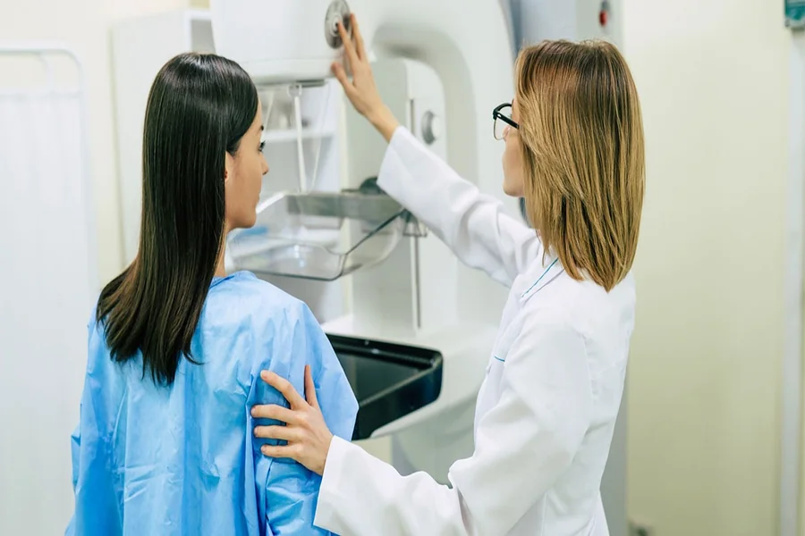
- You stand in front of the machine. A staff member helps you place one breast on a small plate.
- A clear plate comes down to hold the breast still. This can feel tight. It should not feel sharp.
- The X-ray arm moves in an arc over the breast. It takes many pictures in a few seconds.
- The plates release. Then the staff member repeats the process for other views and the other breast.
The machine does not store your touch or voice. It only records images of the tissue. The computer receives the images right away. Then it starts to rebuild them into thin slices.
How the machine creates 3D images
Inside the unit, several parts work together.
- The X-ray tube sends a small burst of energy through the breast.
- Digital sensors on the other side capture that energy and turn it into data.
- Motion controls move the tube in a smooth arc to get many angles.
- Software takes the raw data and builds a set of thin slices through the breast.
The result is a stack of images that the doctor can scroll through on a screen. Each slice shows a different depth. This limits the problem of tissue overlap. With overlap, normal tissue can hide or copy the shape of a problem. With 3D slices, the doctor can see if a spot holds its shape across layers or fades out.
How 3D compares with 2D mammograms
You may wonder if 3D means more radiation or more time. The answer depends on the machine and the setting. Many units now create both 2D and 3D views from the same data. That keeps the dose close to a standard mammogram.
2D vs 3D screening mammograms
| Feature | 2D mammogram | 3D screening mammogram |
|---|---|---|
| Type of image | Two flat pictures of each breast | Stack of thin slices through each breast |
| View of dense tissue | More tissue overlap | Less overlap and clearer layers |
| Chance of call backs for more pictures | Higher | Lower for many people |
| Radiation dose | Standard low dose | Low dose. Often close to 2D when using newer systems |
| Reading time for doctor | Short | Longer because of more images |
You can read more about 3D mammograms on the National Cancer Institute mammogram fact sheet.
Why 3D can mean fewer repeat visits
Many people fear a call back after a mammogram. A call back does not mean cancer. It means the doctor needs more views. With 2D images, folds of tissue or shadows can look like a mass. With 3D slices, the doctor can check if a spot is only overlapping tissue.
This can lead to three gains.
- Fewer call backs for extra pictures.
- Clearer size and shape of any real lump.
- More steady tracking of changes over time.
For some people, this means less worry and fewer visits. For others, it means a real problem is found earlier, when treatment choices are stronger.
What the doctor looks for in the images
The doctor studies each breast in a planned way. You do not see that work, but it follows a clear method.
- The doctor checks the skin line and the nipple.
- The doctor scans each slice for bright spots, lines, or shapes that stand out.
- The doctor compares both breasts side by side.
- The doctor looks at past images, if they exist, to spot change.
3D slices can help separate harmless cysts or normal patterns from signs that need more tests. Those tests might be more views, an ultrasound, or a biopsy. A biopsy is the only way to confirm cancer. The mammogram is the early warning tool.
Safety, comfort, and what you can control
You can control several parts of the visit.
- Tell the staff if you had pain with past mammograms.
- Ask for clear steps before each image.
- Ask how long the compression will last each time.
Compression spreads the tissue so the machine can use a lower dose and still show clear detail. You can ask the staff to pause between views if you need a moment. You can also ask if the unit uses 3D only, 2D only, or both.
The Food and Drug Administration sets quality rules for all mammography units in the United States. You can learn about these rules on the FDA Mammography Quality Standards Act page.
How to use this knowledge at your next visit
Information can cut fear. You now know how the machine moves, how the images form, and why 3D slices can help. You can use that knowledge in three ways.
- Ask if 3D screening is offered and if it is right for you.
- Share any past breast surgery, implants, or strong family history.
- Plan regular screening based on your age and risk, not on fear.
Your body deserves early warning when something changes. 3D screening mammograms are tools to give you that warning with more clarity and fewer false alarms. You stay at the center of each choice.